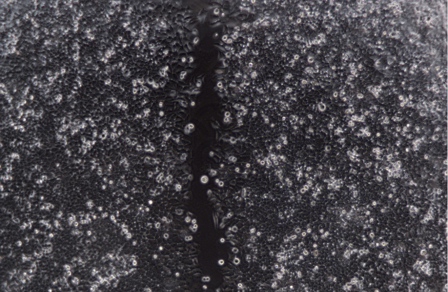
Microscope has become a useful tool in various research fields since its birth and development, providing powerful assistance for disease research, material testing, etc. Specimens are not always easy to see under a microscope, low contrast specimens such as unstained bacteria, thin tissue sections, and living cells.

Live cells without dye labeling have high transparency, strong refraction, and unclear outlines. It is difficult to distinguish them from the culture medium by conventional bright field observation.
For industrial production such as vaccines, monoclonal antibodies, genetically recombined drugs, etc., large-scale cultivation such as cell factories is required. Due to the limitation of the size and height of the container, the microscope is required to have a larger stage and working space.
Cell biology imaging has the following difficulties:
1) The activity and health of cells need the support of suitable temperature, humidity and gas culture conditions. Without these conditions, cells cannot maintain activity for a long time.
2) Unsuitable light sources may damage cell viability and health. High-energy excitation light, high-intensity light, and long-term exposure will all affect the viability and health of cells.
3) Fluorescent dyes may damage cell health. The toxicity of fluorescent dyes and the phototoxicity of the excitation light can lead to poor cell health. Typical examples are DAPI and Hoechst. These dyes are somewhat toxic and are excited with phototoxic UV light.
4) Improper matching of fluorescent dyes will lead to unsatisfactory imaging. Inappropriate or too many fluorescent dyes will lead to spectral overlap, non-specific staining, background signal will be enhanced, and the fluorescence imaging effect will be affected.
5) Fluorescence imaging tests the sensitivity of the camera. Fluorescence imaging belongs to low-light imaging. Ordinary microscope cameras may have the problems of long imaging time and high background noise, and the captured pictures are easy to blur.
6) The focal plane drifts, and the observation target may be out of focus due to movement during the experiment.
The microscope robot independently developed by Bogong Technology can obtain high-definition cell and biological images in real time, and record relevant information including image, activity, and quantity during cell growth. Researchers can observe cell morphology in real time, collect cell images, and perform quality control on the cell culture process to ensure the consistency of product quality.
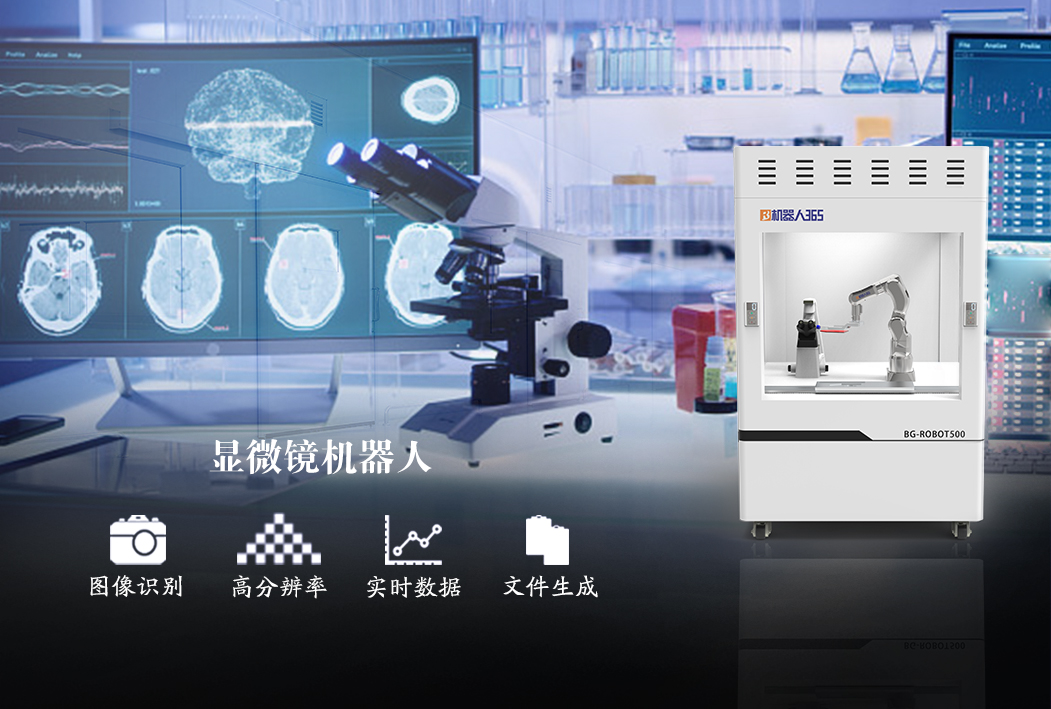
The product is mainly composed of a six-axis robotic arm and an intelligent microscope. It is equipped with an AI cell analysis system, which can flexibly expand a variety of robot applications, support the grasping of various culture bottles, automatically adjust the angle to observe cell morphology, and continuously image and sample for 24 hours.

Product Structure
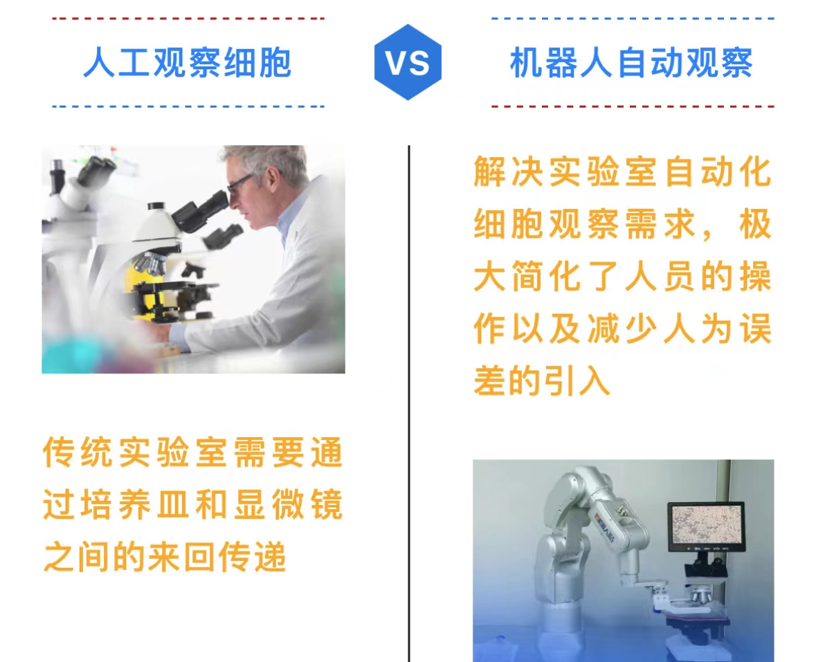
The whole process is completed by machines, and the automatic standardized detection greatly simplifies the operation of personnel and reduces the introduction of human errors. Fast sample processing speed, complete the preparation and detection of a single sample within one minute. Three-axis high-precision electric stage, repeat positioning accuracy of 1μm, precise positioning and automatic focusing.
The microscope online inspection system has been trained by tens of thousands of samples, and uses artificial intelligence technology to perform image recognition. It can record the data completely without setting the recognition parameters, thereby avoiding the difference in results caused by subjective factors.
Product advantages
1. The small footprint makes it possible to observe the microscope in a small space

Microscope robots with higher image quality and ergonomic design can provide stable performance and comfortable workflow for various cell culture needs including live cell observation, cell sampling and processing, image acquisition and fluorescence observation.
2. Simple living cell observation - integrated phase contrast fast cell observation

High contrast Enables clear cell observation at 4x, 10x, 20x and 40x magnifications without changing or re-centering the ring diaphragm, this phase contrast system facilitates simple and efficient cell observation for a faster and more comfortable workflow .
3. Efficient cell observation and processing - compatible with various cell culture containers
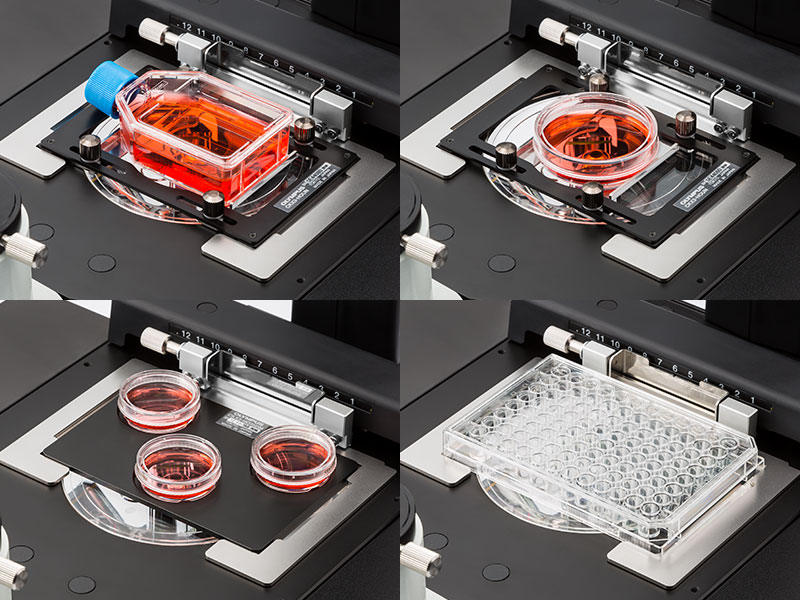
Universal adapter for easy viewing of cells cultured in a variety of containers, including dishes, microplates, and flasks
Optional adapter allows three 35 mm Petri dishes to be mounted on the stage
Holds various types of microplates without adapters
4. Powerful software functions, intuitive software interface
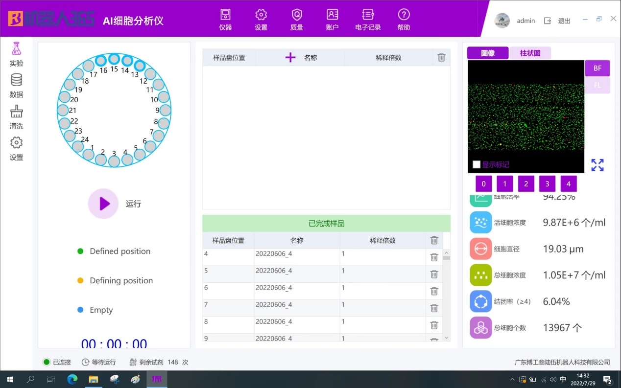
The microscope online detection system has an intuitive operation interface, which is very easy to use and operate. After putting the sample into the microscope, click the corresponding turntable position on the software, and click "Run" to start the detection.
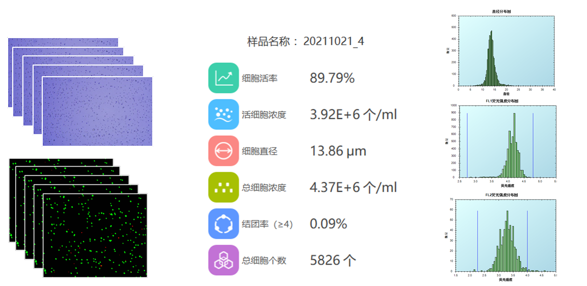
The system has a variety of data display forms, including pictures, diameter distribution diagrams, fluorescence intensity distribution diagrams, etc., from which the operator can learn more about the cells in culture, and can also analyze the state of the cells through the histogram distribution diagram.
